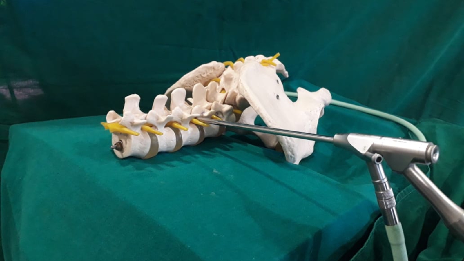
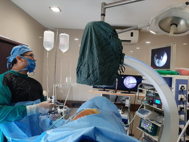
Live Endoscopic Discectomy Procedure In OT

Procedure done under live x-ray guidance
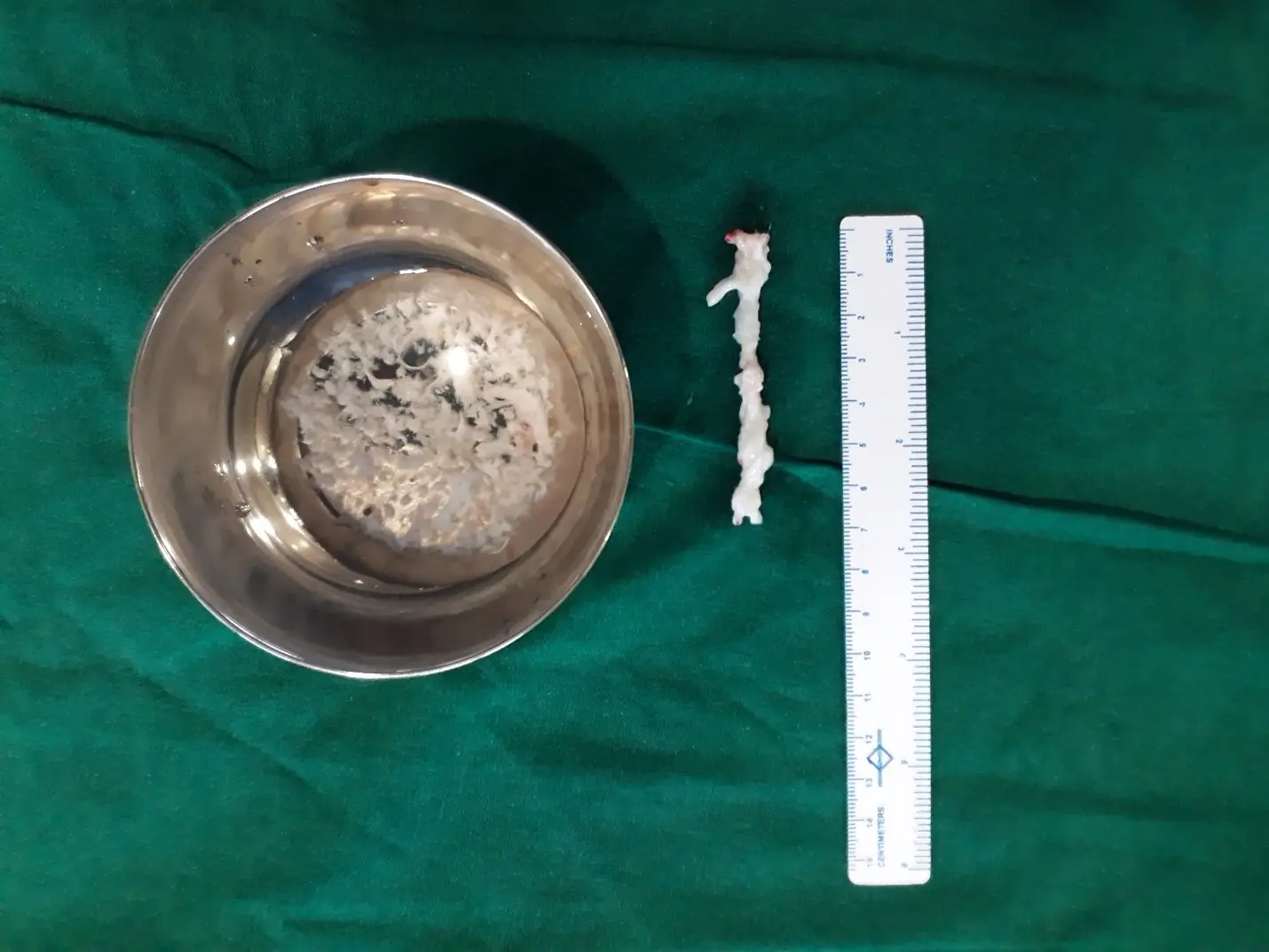
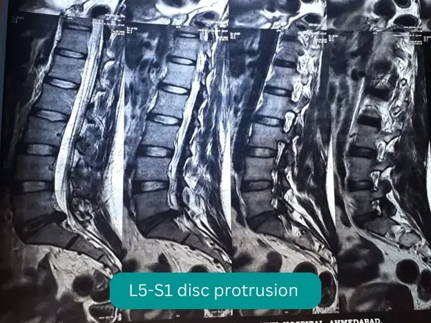
Removed fragments from disc